Skin Fluke
Skin fluke, Gyrodactylus, are common parasites found on the skin and gills of fish. Skin fluke along with gill and eye fluke make up a group of parasitic flatworms known as Monogeneans. There are approximately 3000 species of monogeneans capable of infecting both marine and freshwater species of fish but thankfully the majority of species are host specific meaning they only infect specific species of fish each. This article takes a closer look at skin fluke found on koi and goldfish as this is one of the most commonly identified problems diagnosed at general health checks.
Pathology
Skin fluke predominantly cause damage to fish by attaching themselves to the skin and gill tissue using specialised hooks called ‘haptors’ or ‘anchors’. Parasite attachment sites cause local irritation resulting in inflammation in the area. This is often seen clinically as dulling of colour from excess mucous production or red and inflamed skin. Other symptoms to look out for include clamped fins and ‘flashing’ where fish rubs themselves on the sides or bottom of the tank or pond. Sometimes abnormal jumping behaviour can be seen.
In severe cases, skin fluke infections can lead to ulcer formation. This is due to direct damage at the skin surface and subsequent infection by opportunistic bacteria, which once set in causes erosion and ulceration to the skin.
Parasite attachment in the gills can be result in heavy mucous production which is often seen as white or grey patches or a dull colour to the gill filaments. Unfortunately, heavy skin fluke infections on the gills forms a significant threat to life as extensive damage to the gills will result in poor gas exchange and laboured breathing. In the wrong conditions (warm water temperature and low dissolved oxygen) death from suffocation is a possible outcome in these cases.
Life Cycle
Skin fluke are ‘viviparous’ parasites meaning they give birth to live young, larvae. This is pertinent as some fluke species, especially viviparous species, are capable of doubling in population every 24 hours!
What might have started as a small infection with one or two skin fluke found on skin scrapes can rapidly become a major problem. This is a risk during the spring and summer months as increasing water temperatures directly increases the speed at which fluke reproduce.
Diagnosis
Diagnosis of skin fluke can be made easily via microscopic examination of skin scrapes and gill clips. Although the symptoms listed above can give you a good indication as to whether a skin fluke infection may be present, there are many other parasites that cause the exact same symptoms so microscopy is required to confirm what parasites are actually present.
To diagnose skin fluke at home you will need a microscope, microscope slides and cover slips.
It is recommended to perform skin scrapes with the fish under a gentle sedation in order to keep stress to a minimum and to allow you to collect a scrape from underneath the pectoral fins.
One common misconception is the thought that using a sedative agent like ‘Koi Sedate’ or clove oil will kill or sedate parasites so that when you come to look at your sample under the microscope the parasites won’t be there. This is false, even when using much higher doses of anaesthetic agents, parasites if present, will be seen.
If you are suspicious you have found a skin fluke under the microscope, there are several useful characteristics to look for to confirm this.
- Movement: Skin fluke will shorten and then elongate themselves lengthways, like an accordion, to move across the microscope slide.
- Look for a larvae within the adult fluke.
This is most easily done by looking for haptors within the body of the adult as these are the haptors of the developing larvae.
- Look for the haptors or Opisthohaptors (venus fly trap like appendage, in which the haptors sit) at the end of the fluke.
- V-Shaped head which is often very mobile.

Image 1.1. Skin fluke under the microscope.
Magnification x 100.
a) Circles the attachment end of the skin fluke where the haptors/anchors sit within the opisthohaptor.
b) Larvae within adult two skin fluke.
c) V-shaped heads of skin fluke.
It is also important to appreciate the fact that skin fluke, although being called skin fluke, can also be found on the gills and vice versa for gill fluke, Dactylogyrus sp. So don’t automatically assume a fluke is a skin fluke if found on a scrape of the skin, it could very easily be a gill fluke. There are several ways to differentiate between a skin fluke and gill fluke and these include;
- Gill fluke lay eggs so you won’t find a larvae inside an adult.
- Gill fluke lack the distinctive opisthohaptor and haptors that skin fluke possess.
- Gill fluke have 4 dense, black eye spots at the head end of the fluke, skin fluke do not.
Medical Treatment
The main reason it is useful to be able to differentiate between skin fluke and gill fluke under the microscope is that it will determine what treatment protocol to use.
Unlike skin fluke, gill fluke are ‘oviparous’ which means they lay eggs, this means you will need to use multiple rounds of treatment to treat an infection of gill fluke. This is because the eggs laid by the adults are resistant to treatment, so you need to wait for those eggs to hatch before you can kill them off with treatment. This is achieved through applying multiple rounds of treatment at certain intervals to catch the newly hatched eggs. For most fluke medications repetitive treatments 1 week apart is usually effective in killing gill fluke. Some longer acting medications like FlukeSolve can be repeated every 3 weeks.
Thankfully skin fluke are viviparous and do not lay eggs so in theory, one effective round of treatment should kill off the entire life cycle, larvae and all.
Unfortunately treating skin fluke isn’t always as straight forward as you would hope. Over many years, fluke have developed resistance to many of the common fluke medications available on the market. I have heard some clients describe fluke as ‘bomb proof’ and in some cases I wouldn’t disagree! Unfortunately there is no way to predict what fluke treatment will work best for a pond and it may take a bit of trial and error to find the right product. For the best chance of a successful treatment outcome, ensure you are dosing accurately by calculating the exact volume of your pond first. If you have an irregular shaped pond, you can use salt and a salt meter to help you calculate the exact volume. Please email for guidance on this.
London Aquatic Veterinary Services will usually always attempt to treat all fluke infections whether skin and gill fluke, with prolonged salt therapy first. The standard salt therapy course involves holding the fish at 0.5% (5g/L) salinity for 4 weeks. It is crucial that the salt level of the water does not dip below 0.5% for the entirety of the course to ensure maximal treatment success. This therefore involves routine salinity testing with a saltmeter and topping up on salt when any water changes are performed. If after repeat skin scrapes the fluke infection is still present, an over-the-counter medication usually containing praziquantel will be opted for.
In some cases, pre-treating the fish with salt or Chloramine T can reduce the mucous layer on the fish to allow for better penetrance of the fluke medication when used.
Please note! Exercise care when using salt. There are many over-the-counter medications that are toxic when used in combination with salt such as malachite green and potassium permanganate formulations.
Once salt is in a tank or pond, it will take a long time to come out so take care if using medications shortly after salt therapy and seek guidance if unsure and always use a salt meter.
Non-Medical Control
Aside from the various medical options for treating skin fluke there are several non-medicinal action points you can take to reduce the risk of a skin fluke outbreak;
- Maintain excellent water quality.
- Keep organic loading to a minimum by routinely siphoning or vacuuming the bottom of the tank or pond.
- Quarantine all new fish and scrape the skin to check for parasites before adding them to the main pond or tank. If an infection is present, treat the fish in quarantine first before adding them to intended pond or tank.
In rare cases, some infections can never fully be cleared. In these instances, reducing the number of skin fluke rather than total eradication is the aim. In these cases following the non-medicinal control points highlighted in the paragraph above will aid this process.
Prognosis
Once identified, skin fluke infections usually carry a very good prognosis, in fact, the identification of one or two rogue skin fluke may not warrant any medical treatment at all. As a rough guide, if clinical symptoms such as flashing or clamped fins or excess mucous are seen along with skin fluke on scrapes, then treatment is warranted. If only a handful of fluke are found on scrapes, the water temperature is low or the fish aren’t displaying any clinical symptoms then either treatment can be delayed until the spring or not used at all.
If treatment is deemed necessary, appropriate medication and protocols should clear most ponds or tanks of an infection relatively easily. Unfortunately, there are some nightmare cases where skin fluke populations appear to be multi-resistant to various medications. Should resistance be suspected or you would like help diagnosing skin fluke in your pond, reach out to London Aquatic Veterinary Services for guidance.
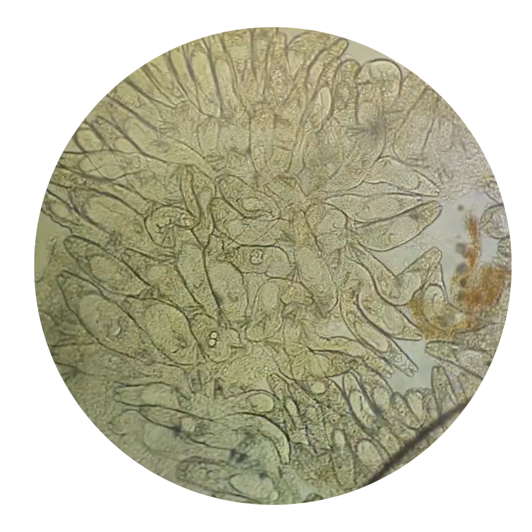
Image 1.2 Severe skin fluke infection on a goldfish skin scrape.