Dropsy
Dropsy, one of the most feared terms amongst hobbyists and aquarists alike but what does it actually mean? First and foremost, dropsy itself is not a specific disease but a set of clinical symptoms, known as a syndrome. The clinical symptoms which make up this syndrome include swelling of the body cavity or coelom as well as the appearance of lifted scales colloquially known as ‘pine coning’ and exophthalmos (bulging eyes). All symptoms can occur simultaneously or in isolation.
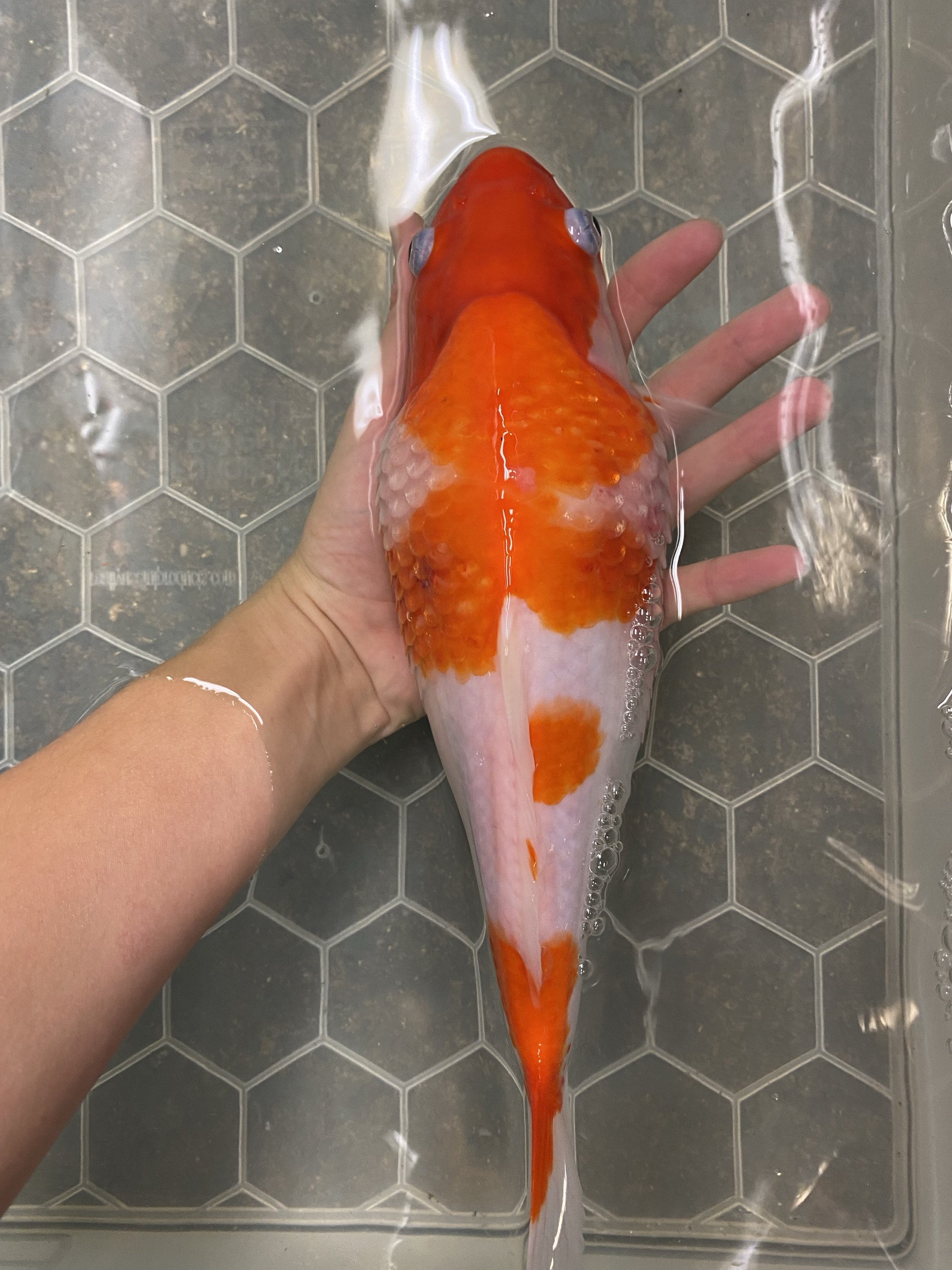
Dropsy can essentially be thought of as oedema, which is the scientific name for a build-up of fluid within bodily tissues. The classic pine cone appearance associated with dropsy occurs due to scales being pushed out from their normal alignment due to fluid building up within the skin in between the scales. Similarly, exophthalmos is caused by fluid building up behind the eyes. Changes can be subtle at first so viewing the fish from above can help where it is easier to appreciate the fish’s contour and shape.
Unfortunately, once dropsy is present the prognosis is guarded as its presence often signifies advanced and severe internal disease.
The kidneys of fish
The fish kidney, as with all vertebrate species, governs fluid and water balance within the body. The kidneys are adapted to deal with one of the most important physiological challenges acting on aquatic organisms, osmosis, the movement of water across a semipermeable membrane.
Following the law of osmosis, water molecules move from areas of high concentration to areas of low concentration. So for a fish in a freshwater environment, there is a higher concentration of water molecules surrounding the fish when compared to inside the tissues of the fish. Following the law of osmosis, water is moves into the tissues of the fish, from an area of high concentration of water molecules to an area of low concentration. Uncontrolled, water would continue to move into the fish and cause the fish the swell up with fluid. Thankfully, the kidneys of freshwater fish are perfectly adapted to this constant movement of water into the tissues by producing high volumes of dilute urine to remove excess fluid. It makes sense then, that when the kidneys stop working properly, fluid starts to build up in the body of the fish, resulting in the symptoms that make up dropsy – lifted scales, free fluid in the coelom and exophthalmos.
Therefore, when dropsy is present we can deduce that there is some level of kidney disease. The trick to treating dropsy effectively is to work out what is causing the primary damage to the kidneys in the first place. Damage to the kidneys can be genetic, infectious or neoplastic (tumorous) in origin and looking at the wider picture, influenced by poor water quality and husbandry.
It is important to understand that the fish kidney, as with other vertebrate species, has a very low regenerative capacity, once damaged, the damage is irreversible.
Causes
-
One of the most common genetic disorders affecting the kidneys of freshwater fish, especially goldfish, is Polycystic Kidney Disease (PKD). Frustratingly, being a genetic condition, there is nothing that can be done to prevent or treat this disease. PKD is a degenerative disease with symptoms of dropsy being slow to develop over time.
Coelomic swelling can be massive, with some fish swelling to almost twice their normal size. Cases can deteriorate to such a stage where fish are unable to swim around efficiently or eat and in extreme cases thinning of the body wall can be seen. Coelomic swelling is caused by numerous expanding, fluid filled cysts within the kidney tissue. Gradually healthy kidney tissue is destroyed, reducing the functional capacity of the kidney to remove excess fluid from the body. Ultrasound is the best diagnostic tool for this disease as it is very easy to see the thin-walled fluid filled cysts.
-
Some causes of coelomic swelling which are not a result of fluid build-up within the body cavity include neoplastic coelomic masses. Gonadal tumours are common amongst koi and goldfish varieties and can be identified via radiography and ultrasonography. It is important to understand that although often affecting organs distant to the kidney, coelomic tumours can cause dropsy via growing and exerting physical pressure on kidneys and reducing the functional capacity indirectly.
Although rarely diagnosed a tumour of the kidney tissues itself can also cause dropsy.
-
Dropsy can be seen with various infectious diseases in fish such as bacterial and viral infections. In some cases other external symptoms such fin rot and ulceration may be present which can aid diagnosis.
Diagnostics
Some of the most useful tests available to fish veterinarians to explore coelomic swelling is the use of diagnostic imaging such radiography (x-rays) and ultrasound. Ultrasound is the preferred modality as it particularly good for visualising fluid.
Treatment
One of the most effective and non-specific treatments for dropsy is dosing the aquatic environment with a high dose of salt. By adding salt to the water, you are adjusting the osmotic balance and reducing the movement of water pulled into the tissues via osmosis. This action alleviates some pressure on the kidneys and can help reduce the severity of fluid retention. You will often hear people say that adding salt ‘reduces stress on the fish’ this is somewhat correct but more specifically it reduces the osmotic stress on the fish.
If an infectious cause behind dropsy is suspected, injectable antibiotics may be opted for. In cases where there are no viable treatment options (PKD, coelomic masses etc) or in cases where the fish is simply too far gone, euthanasia is often recommended.
Dropsy is feared and quite rightly. The presence of lifted scales in most cases indicates severe and irreversible damage to the kidneys. Treatment is tricky but not impossible in all cases, they key to success is to act quickly. London Aquatic Veterinary Services can help identify the likely cause of dropsy via on site physical and ultrasound examination and give guidance on the prognosis and any suitable treatment protocols.